


Our activities focus on developing computational techniques as enabling technologies for tissue engineering. By leveraging advanced modeling and simulation tools, we aim to better understand the complex interactions that govern tissue development and regeneration. Our work includes the development of predictive algorithms to optimize process parameters in the manufacturing process, as well as the simulation of mechanobiological behaviors. These computational solutions help reduce reliance on trial-and-error, streamline experimental workflows, with the aim to ultimately contribute to more efficient and effective tissue engineering strategies.
Extrusion-based bioprinting
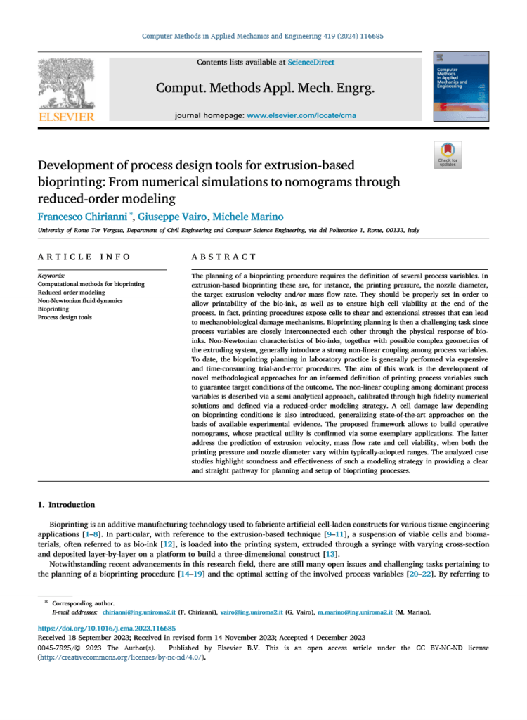
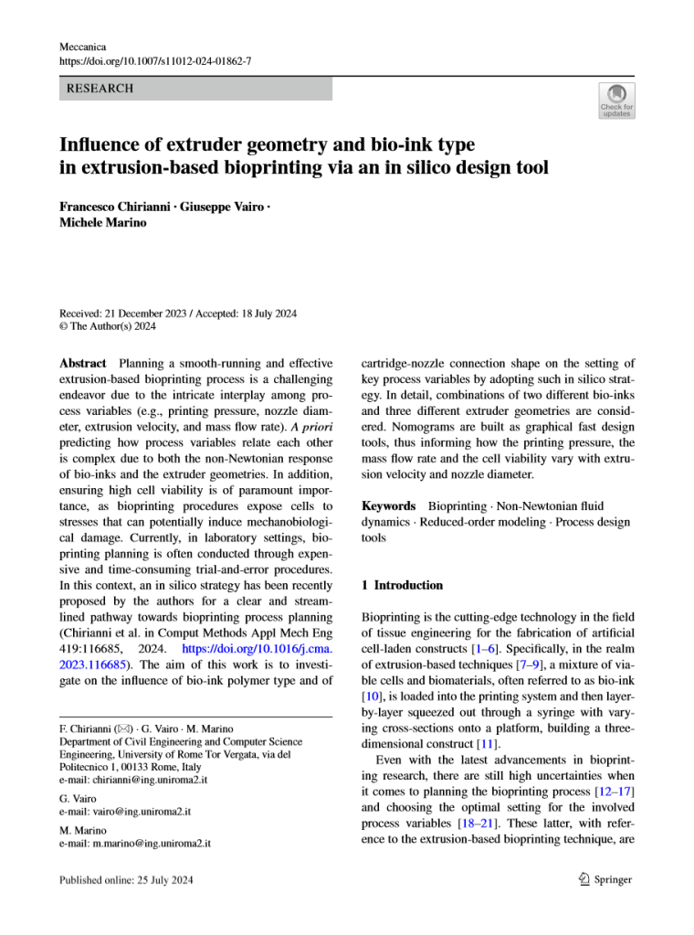
In-silico design tool for the planning of bioprinting processesExtrusion-based bioprinting is the cutting-edge technology in the field of tissue engineering for the fabrication of artificial cell-laden constructs. Planning a smooth-running and effective bioprinting process is a challenging endeavor, since the choice of process variables should fulfill technological demands, as well as ensure the utmost cell viability by the end of the process. To date, the bioprinting planning in laboratory practice is generally performed via expensive and time-consuming trial-and-error procedures.
We have developed an in-silico strategy for an informed definition of printing process variables such to guarantee target conditions of the outcome. This framework allows to build operative nomograms as graphical fast design tools. We have also proposed a cell damage law depending on bioprinting conditions, generalizing state-of-the-art approaches on the basis of available experimental evidence.
Hydrogels properties
Multiphysics theoretical and computational modellingSee our activities on hydrogels modelling
Modelling of neotissue formation
Cell behavior in three-dimensional scaffoldsThe major limitations in the in vitro production of tissues are time and cost, which significantly hinder the scalability of tissue production and, consequently, the broader applicability of tissue engineering technologies. During the first phase of tissue development in hydrogel scaffolds, cell clusters form, serving as the progenitors of tissue blocks. As a result, the mass of the tissue construct that can be produced within a given time is closely linked to cell motility during this initial stage.
Theoretical and computational models for cell motility within polymeric hydrogel scaffolds have been developed and refined over the years. A phase-field model was created to describe cell motion within naturally degrading hydrogels. These simulations capture the early stages of tissue development, including the mutual adhesion and fusion of small cell clusters. The framework has also been extended to account for the effects of cell enzyme release, which continuously degrades the hydrogel, thereby facilitating cell movement and contributing to tissue formation.


Numerical results emphasize the critical interplay between nutrient availability within the construct, chemoattractant and enzyme production, scaffold degradation, and cell motility. The model has shown quantitative agreement with experimental data, accurately capturing characteristic times and cell velocities. These findings highlight the potential of the developed in silico framework as an effective computational tool for optimizing neotissue mass production. By simulating these complex interactions, the model offers valuable insights that could pave the way for more efficient and scalable tissue engineering processes.
The complexity of tissue engineering applications based on bioprinting technologies are described from a multidisciplinary perspective in the book co-edited by our group:
Bioprinting – From Multidisciplinary Design to Emerging Opportunities
Within this book, chapter 6 is dedicated to models and simulations as enabling technologies for bioprinting process design.